WHAT CAUSES THE DISEASE?
What is Aortic Stenosis?
The heart has four valves that ensure the blood circulates in one direction in an orderly fashion throughout the body. The aortic valve sits between the main pumping chamber of the heart (left ventricle) and the main artery of the body (aorta). The average thickness of the aortic valve is only 0.3 mm, yet the aortic valve will open and close nearly 2 billion times in a person’s lifetime, this results in a high probability that the aortic valve will be damaged. In addition, bacterial infection, aging and other factors could also threaten the health of the aortic valve, resulting in aortic stenosis (AS).
When AS occurs, the valve becomes hard, the flexibility is reduced, and cannot be fully opened, affecting the normal pumping mechanism of the heart, resulting in insufficient blood output. In response, the heart has to increase its power to pump out the same amount of blood. Over time, this long-term ‘overworked’ state will permanently damage the heart muscle, causing heart failure. Heart failure can seriously affect patients’ daily activities and even result in death.
Following hypertension and coronary artery disease, AS is the third most common cardiovascular disease, and is highly linked to aging. Research has found that about 7% of the population over 65 has AS, the prevalence of AS for those between 50–59 is 0.2%, 60–69 is 1.3%, 70–79 is 3.9%, 80–89 is 9.8%.


SYMPTOMS OF SEVERE AORTIC STENOSIS
In mild cases, severe aortic stenosis is usually asymptomatic. But as the disease progresses, the pressure needed to pump the blood through the narrow valve increases. The following symptoms could spell danger for patients with severe aortic stenosis:
- Dizziness (or Syncope).
- Shortness of Breath (or Dyspnea).
- Fainting.
- Chest pain (or Angina).
- Fatigue
- Difficulty in exercising or completing daily activities.
Severe aortic stenosis is a serious heart problem, the outlook for patients can be poorer than many cancers that have metastasized (e.g. lung and breast cancer). Without replacing the valve, patients who experience chest pain have an average life expectancy of 5 years, those with fainting an average life expectancy of 3 years, and only 2 years with patients who experience breathing difficulties.
DIAGNOSIS OF SEVERE AORTIC STENOSIS
- Echocardiogram (Echo): Through using ultrasound, this test produces video images of the heart in motion. Doctors can then make a diagnosis based on the observed conditions of the heart and the heart valve.
- Electrocardiogram (ECG): Severe Aortic Stenosis affects the way the heart beats, and enlarges the heart chamber. Through ECG, doctors can make a diagnosis based on detecting the electrical activity of the heart.
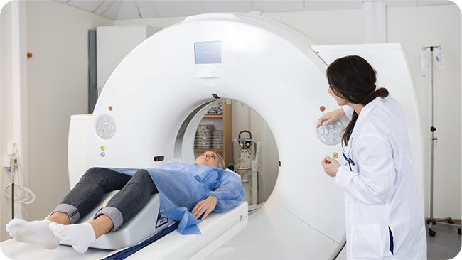
- X-ray or CT scan: Through making single or multiple images of the heart and heart valves, doctors can make a diagnosis based on the enlargement of the heart, the aorta and the
calcium build up in the heart valves. - Other Diagnoses: Other diagnosis methods doctors could use to support their decision for treatment include, exercise or stress tests, MRI, and catheterization.


TREATMENT PROCEDURES
Currently for patients with aortic stenosis, the main treatment options are pharmaceutical or surgical procedures. However, pharmaceuticals are used mainly to slow down the progression of the disease, and cannot completely cure the disease. Therefore, surgical procedures are the only real option for treating aortic stenosis. There are currently two types of surgical treatment for treating this disease, the traditional open-heart surgery or minimally invasive surgery.
Surgical aortic valve replacement (SAVR)
During SAVR, or open-heart surgery, the patient is first anesthetized, then placed on a cardiopulmonary bypass, and put in a state of cardiac arrest. After, a 20cm incision is made on the chest and then the aorta, to access the diseased valve. Finally, the diseased valve is then replaced with an artificial valve. The operation time and post-operative recovery time for this procedure is long. As a result, elderly patients with poor body condition or cardiac function cannot be treated with this operation. In addition, patients may face higher risk of stroke, myocardial infarction, and even death post-surgery.
Transcatheter aortic valve replacement (TAVR)
During TAVR, or minimally invasive surgery, with a small incision in the femoral artery of the thigh, the artificial valve is transported through the catheter to the aortic valve, and released to replace the diseased valve, this restores the normal opening and closing function of the heart ‘valve’, and as a result relieves the signs and symptoms of AS, and improves the overall health and quality of life for patients.